
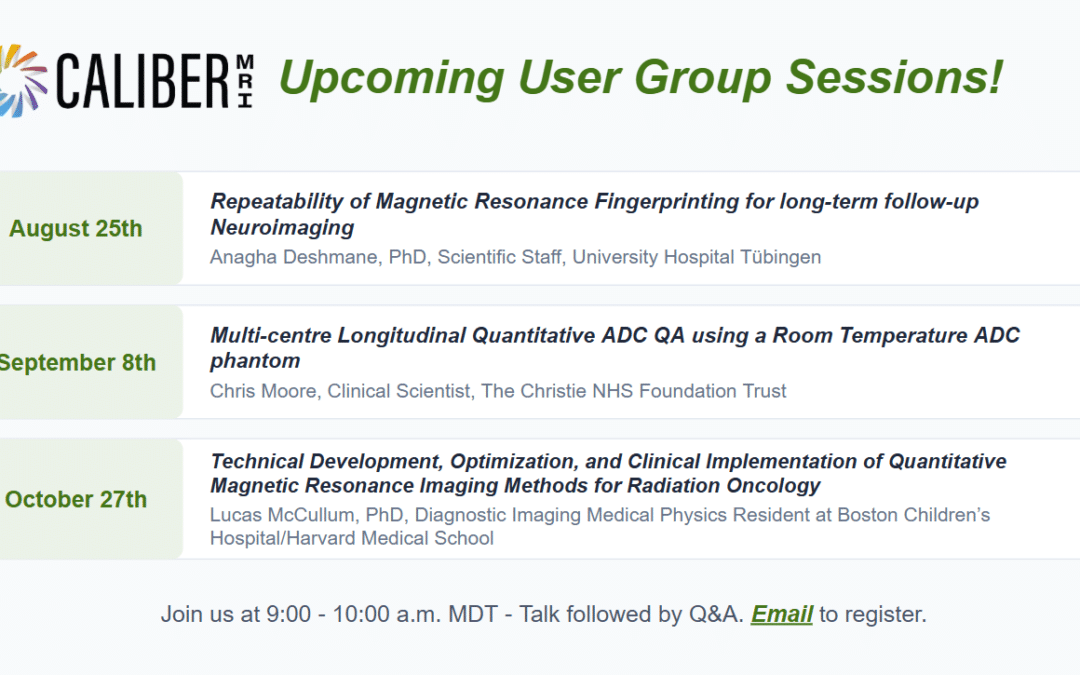
Please join us for our upcoming User Group Sessions!
- August 25th (9:00 a.m. MDT)
Dr. Anagha Deshmane, PhD, University Hospital Tübingen, Tübingen, Germany, will be discussing work presented at the 2026 ISMRM meeting in Cape Town: Repeatability of Magnetic Resonance Fingerprinting for long-term follow-up Neuroimaging.
Dr. Deshmane’s work focuses on the development and clinical translation of new methods with magnetic resonance imaging; quantitative and molecular imaging technologies; Magnetic Resonance Fingerprinting (MRF); and CEST (Chemical Exchange Saturation Transfer). - September 8th (9:00 a.m. MDT)
Title: Multi-centre Longitudinal Quantitative ADC QA using a Room Temperature ADC phantom
Summary: Work spanning 2 years, 9 scanners at 5 centers to evaluate repeatability and reproducibility of quantitative ADC measurements in preparation for longitudinal clinical trials incorporating quantitative ADC as a biomarker of cancer, using the qMRI room temperature ADC phantom. Study results and discussion of the utility of the phantom.
Speaker Bio: Chris Moore – clinical scientist, The Christie NHS Foundation Trust. Chris completed an undergraduate physics degree at The University of Manchester. After a brief time as a software engineer, he successfully applied for the Scientist Training Programme (STP) in medical physics. He completed the programme in September 2021 and has taken on a clinical role in the MR group. Chris splits his time between regional physics support and providing research support to the Radiotherapy Related Research group, with research interests including image registration, quantitative MR imaging biomarkers and longitudinal quantitative quality assurance of DWI images. - October 27th (9:00 a.m. MDT)
Title: Technical Development, Optimization, and Clinical Implementation of Quantitative Magnetic Resonance Imaging Methods for Radiation Oncology
Summary: Magnetic resonance imaging (MRI) has gradually grown more important in the field of radiation oncology. This paradigm shift has grown from initial experiments with MRI of cancerous lesions in the early 1970s, to the integration of MRI images for radiation therapy treatment planning (i.e., MRI-Simulation) in the early 1980s, to the combined MRI linear accelerator (MR-Linac) becoming commercially available in 2018 which can simultaneously image a tumor and treat it with radiation. However, due to its novelty in radiation oncology, its full potential for the integration of quantitative MRI-based biomarkers has yet to be fully realized. Therefore, this talk will provide a framework for this integration from the initial technical developments, to the subsequent optimization, to the ultimate clinical implementation in the radiation oncology workflow. This work will be presented in three distinct aims: 1) the technical development of quantitative MRI techniques in radiation oncology, 2) the technical optimization of these techniques for site-specific applications, and 3) their clinical implementation and potential use cases. Several techniques will be explored for quantitative T1, T2, PD, R2*, proton density fat fraction (PDFF), and triglyceride composition mapping. Most of the techniques presented will acquire multiple of these quantitative parameters simultaneously due to the unique time constraints in radiation oncology, especially on the integrated MR-Linac.
Speaker Bio: Lucas McCullum completed his B.S. in Mechanical Engineering and B.A. in Applied Mathematics at The University of Maryland – Baltimore County in 2019. Following this, he worked as a research software engineer at the Massachusetts Institute of Technology in the Laboratory for Computational Physiology under the advisership of Dr. Roger Mark M.D., Ph.D. developing open-source tools and databases for health informatics. He then worked as a research assistant at Massachusetts General Hospital in the laboratory of Dr. Harald Paganetti Ph.D. and Dr. Clemens Grassberger Ph.D. focusing on computational simulations of lymphocyte depletion during radiation therapy. In 2022, he started his Ph.D. in Medical Physics at The University of Texas MD Anderson Cancer Center Graduate School of Biomedical Sciences under the advisership of Dr. Clifton D. Fuller M.D., Ph.D. where he developed and clinically implemented quantitative magnetic resonance imaging techniques in the radiation oncology department and successfully defended his Ph.D. in 2026. He is currently a medical physics resident in diagnostic imaging and nuclear medicine at Boston Children’s Hospital and Harvard Medical School.
Email us here if you’d like to attend the sessions! Format: One hour call – Presentation followed by Q&A.